Available measurement techniques:
- Bragg –Brentano standard powder diffraction (BB,XRD) – crystal phase analysis
- Grazing Incidence X-ray Diffraction (GIXRD) – diffraction at fixed small incidence angle suitable for thin films
- In-plane Diffraction (also abbreviated GID: grazing incidence diffraction) – diffraction of planes (almost) perependicular to the sample surface, limited by primary beam intensity
- X-ray Reflectivity (XRR) -optical specular/diffuse reflection – thin films thickness, density, roughness of interfaces
- Small angle X-ray scattering (SAXS) (only for suitable samples, limited by primary beam divergence and intensity) size, orientation, shape and composition of nanoparticles – transmission/reflection mode
- High resolution XRD (HRXRD) – rocking curves, reciprocal space mapping – epitaxial layers, lattice misfit, relaxation, diffuse scattering on defects
Texture, Strain & Stress, Small area analysis (<1mm2) -orientation distribution function, epitaxial relationship - All the above methods in-situ at non-ambient temperature up to 1100°C (in vacuum >0.1Pa, inert gas, air)
Equipment:
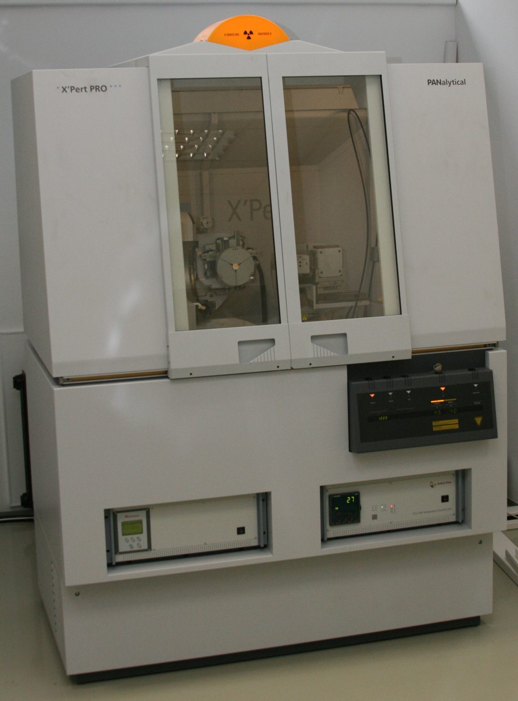
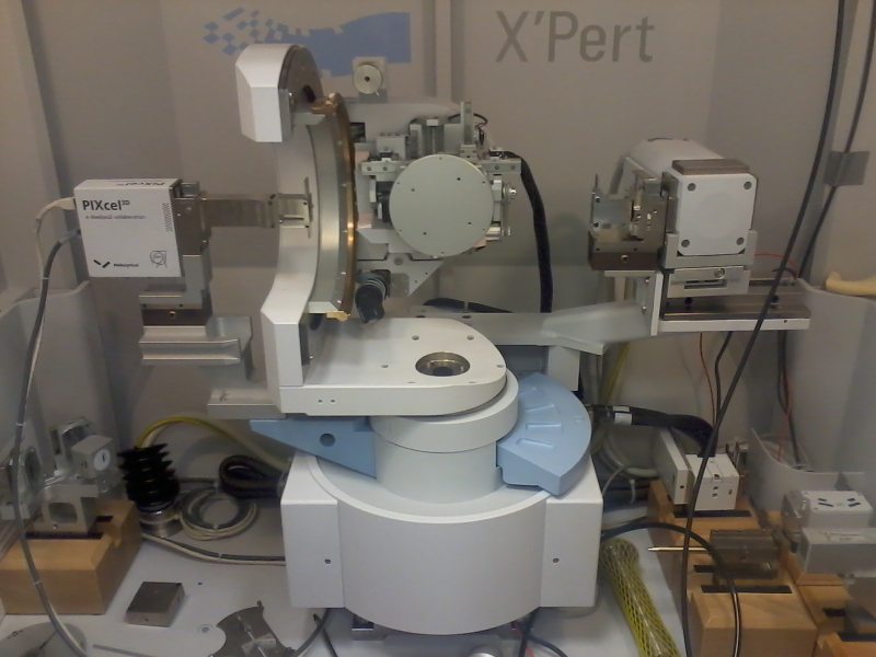
PANalytical X’Pert PRO MRD diffraction system with static CuKa X-ray tube; horizontal high resolution goniometer; 5-axis Euler cradle; modular PreFIX X-ray optical elements; easily changeable line / point focus; PIXcel 3D solid state area detector (255×255 pixels, 14x14mm, linearity: 50 kcps/pixel, 12.8 Mcps/strip); Sample size: diameter < 100mm, thickness < 15mm , sample weight < 1kg.
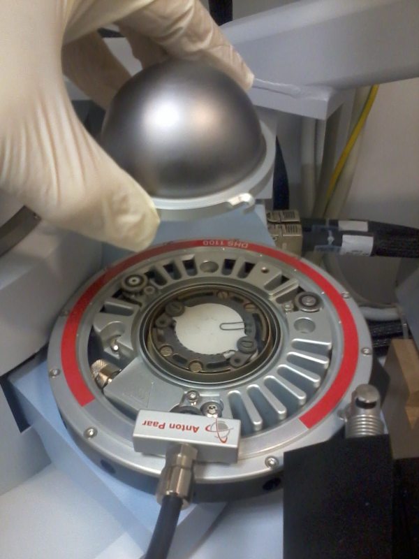
Non-ambient high temperature chamber type Anton Paar DHS 1100 with domed heated stage; range: RT..1100°C; cooling via pressed dry air; geometry of holder with graphite dome allows usage of Euler cradle; at normal pressure or rough vacuum, inert gasses, nitrogen, oxygen, air; ramp up max. 500°C/min; Sample size: diameter < 20mm, thickness < 2.4mm
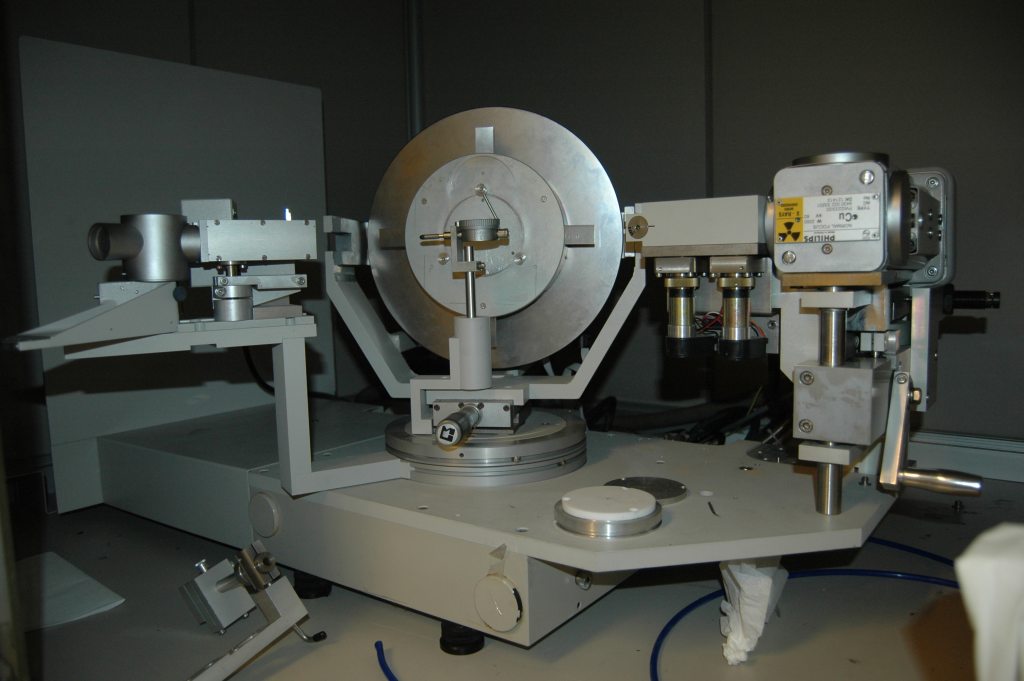
2nd diffraction system: PHILIPS Material Research Diffractometer; HRXRD/RSM, XRR: Adjustable four-crystal monochromator Ge220/Ge440; Triple axis /Receiving slit module; XRD with suppressed fluorescence: Cross-slit collimator, Parallel plate collimator + diffracted beam monochromator
Typical results
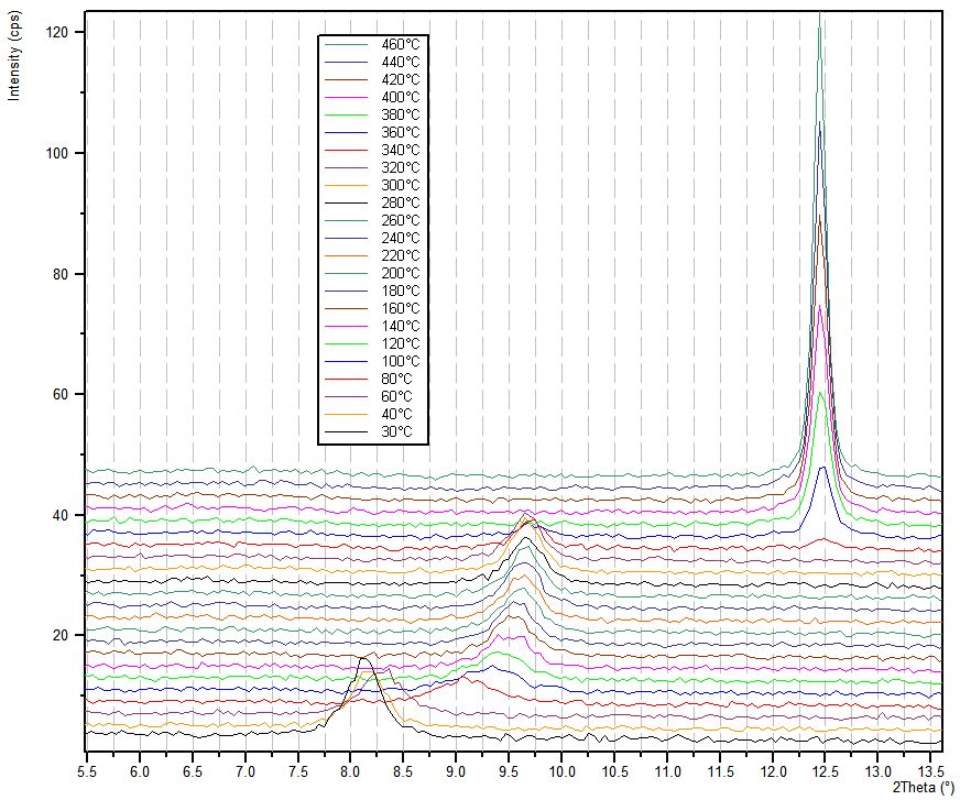
In-situ study of recrystalization in vanadium oxide layer deposited by sol-gel method on glass substrate.
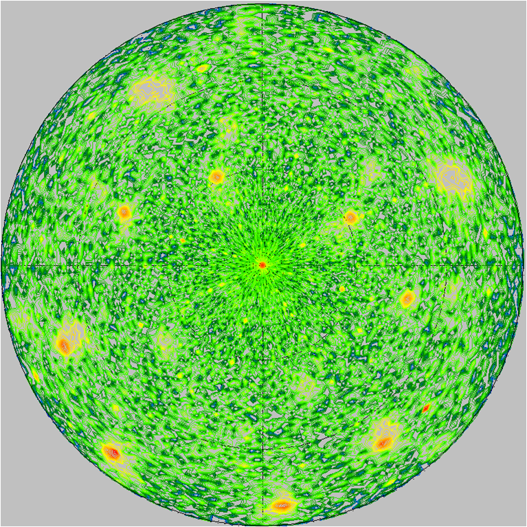
Texture pole figure measured in 211 diffraction position of TiO2-Rutile layer sputtered on r-cut Al2O3.
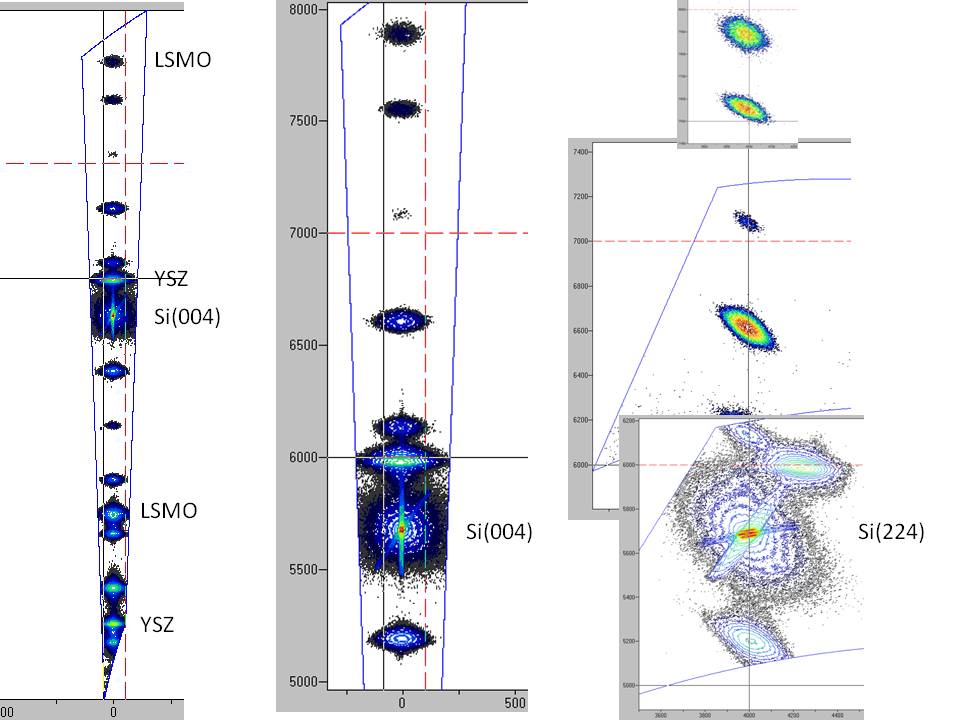
Example of high resolution reciprocal space maps of epitaxial LSMO/BTO/YSZ/Si/SiO2/Si.
Staff:
doc. Dr.techn. RNDr. Tomáš Roch
e-mail: roch@fmph.uniba.sk, Phone: +421-2-60295-270, 189